
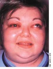


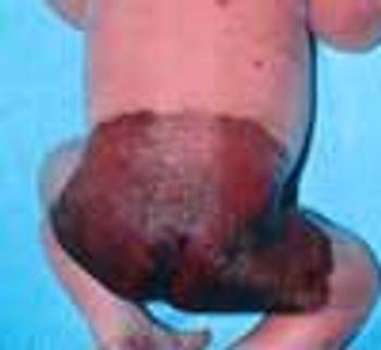
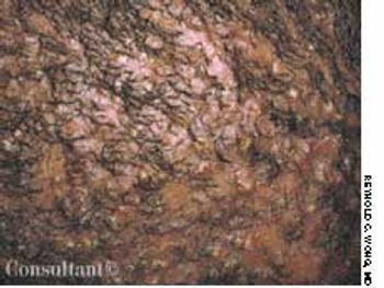
This is a congenital melanocytic lesion (also known as giant hairy nevus or giant congenital pigmented nevus).
This is a congenital melanocytic lesion (also known as giant hairy nevus or giant congenital pigmented nevus).
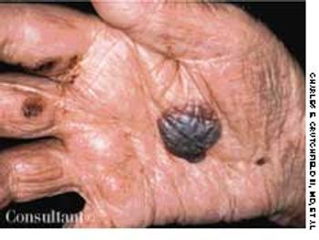
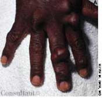
This lesion erupted on a 58-year-old man's right palm, and several tense bullae also were visible on the trunk. The patient complained of mild pruritus. He had no history of similar lesions. A routine skin biopsy was performed, and the diagnosis of hemorrhagic bullous pemphigoid was made.
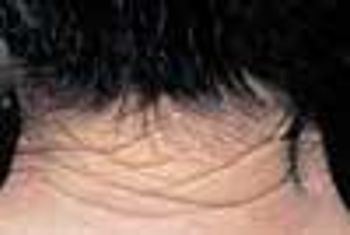
During a routine skin cancer screening, yellowed, thickened, leathery skin was noted on the posterior neck of a 73-year-old retired construction worker. Colloquially, this condition is called “sailor's skin” or “farmer's skin” and is seen in persons who have had long-term exposure to the sun. It is known clinically as cutis rhomboidalis nuchae, because the well-defined furrows in the skin resemble an irregular rhomboidal pattern.
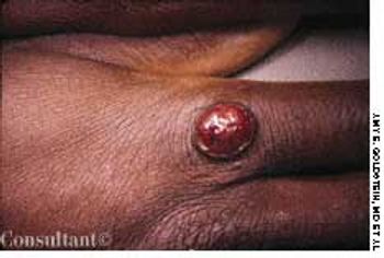
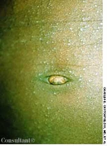
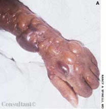
A 32-year-old man who had no significant medical history complained of “something growing on the knuckles of my right hand.” He reported that a “bump” was forming on the site of a cut he sustained while slaughtering sheep 3 weeks earlier. There was no blister, discharge, or pain. The patient denied any fever, cough, or malaise. He also did not recall seeing any lesions or “bumps” on the sheep.
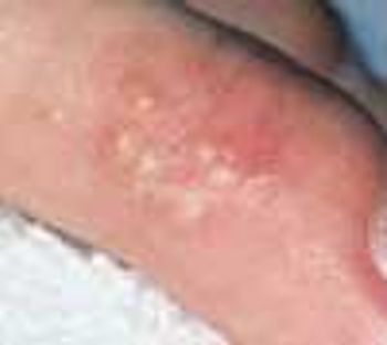
A 40-year-old dental assistant requested a prescription for antibiotics to treat the acute outbreak of painful, deep blisters that had recurred on her index finger. Prior eruptions of similar lesions had been diagnosed as staphylococcal infections and were treated with antibiotics.
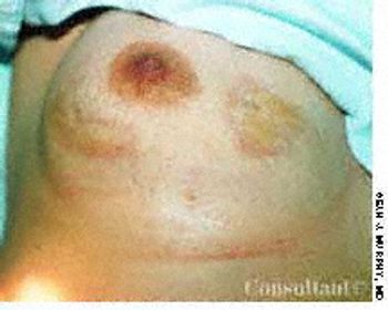
Two enlarging, dry, tender lesions had developed on the right breast of a 62-year-old woman 2 years before she sought medical consultation. The patient had no other symptoms; she was taking metoprolol succinate for cardiac arrhythmias.
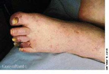
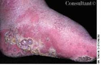
A 65-year-old man presented with bluish toes and petechiae on his toes and feet 3 days after having undergone renal angiography. Bilateral pitting pedal edema was noted, and laboratory tests revealed proteinuria, eosinophilia, and an erythrocyte sedimentation rate (ESR) of 65 mm/h.
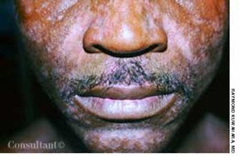
This 27-year-old man complained that a facial rash of several years' duration had worsened during the past few months. Hypopigmented macules with scale were especially prominent on the eyebrows and in the nasolabial folds; a moderate amount of scale was noted on the scalp. The patient was seropositive for HIV.
A 56-year-old man was admitted to the hospital with right lower lobe pneumonia, which was exacerbated by smoking-induced chronic obstructive pulmonary disease (COPD).
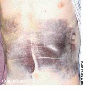
An 80-year-old man with a history of congestive heart failure, coronary artery disease, cardiomyopathy, and thoracic and abdominal aneurysms was taken to the emergency department because of mental status changes, back pain, and ecchymotic areas over his body. The ecchymoses started on his back 5 days before admission and spread to his abdomen.
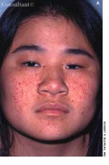
Tan-pink acneiform lesions on the face of a 15-year-old girl had not responded to topical acne therapy. A 1 × 0.5-cm, elevated subcutaneous nodule was noted on the right lateral knee. The lesions on her face and knee had been present for 11 years. The family history was noncontributory.
An otherwise healthy 4-year-old boy was brought for evaluation of a mildly pruritic rash, which had been present for approximately 8 months. The developmental history of the eruption was equivocal, and the child's mother reported no aggravating or ameliorating factors.
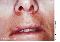
The tender, crusted nodule on the lower lip of a 35-year-old man had been growing slowly for 6 months. The patient reported a history of extensive sun exposure.
A 35-year-old man had a 5-year history of progressive hair loss characterized by follicular inflammation with destruction of the follicle and consequent permanent alopecia. Almost the entire scalp was involved. A few pustules were seen on examination, but the clinical picture was mostly one of scarring and irreversible hair loss.
These benign vascular tumors are sometimes seen in pregnancy.
A 69-year-old man with a long history of lymphedema secondary to repeated cellulitis sought medical care for mildly pruritic, nontender, purple nodules that had erupted on the bottom and side of one foot 6 months earlier. Scale surrounded the nodules.
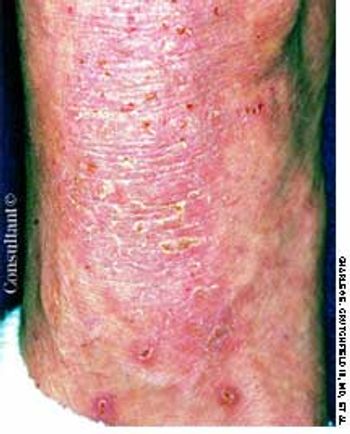
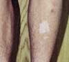
A 41-year-old man sought treatment of the rough, intensely itchy patch on the dorsum of his left foot and lateral ankle. There were no other similar plaques on his body.
A 79-year-old nursing home resident was hospitalized for evaluation of hyperkalemia and leukocytosis. Her medical history included hypertension, respiratory failure with subsequent tracheostomy placement and ventilator dependency, and anemia. Both of her legs had been amputated above the knee secondary to complications of type 2 diabetes mellitus.
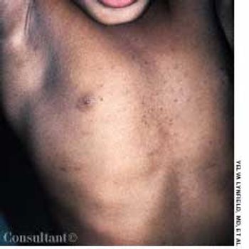
Asymptomatic flesh-colored papules were noted on the chest of a healthy 4-year-old black girl. Some of the 1- to 2-mm papules had central, comedo-like crusts. The remainder of the skin was normal.
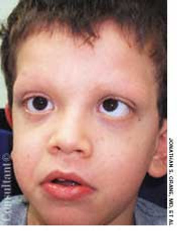
The mother of a 7-year-old boy with cardiofaciocutaneous syndrome sought treatment for the cutaneous aspects of her son's disease. Dry skin and keratosis pilaris of the upper outer arms were noted.
A 17-year-old high school athlete was anxious about this new “mole” that appeared on the heel of his right foot. His concern was prompted by the recent diagnosis of melanoma in his aunt.

After 3 months of seeing this painless mass at the angle of the 3-year-old's left jaw, his parents sought medical advice for their son. The youngster had no constitutional symptoms. A Mantoux test was performed, and an erythematous, indurated area measuring 15 mm in diameter was found at the test site 48 hours later.

Concerned about a lesion between her eyes, a 91-year-old woman sought medical evaluation. She had not seen a physician for 23 years.
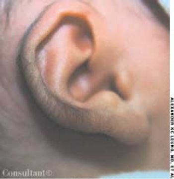
A boy was born to a gravida 2, para 1, 26-year-old woman at 37 weeks' gestation. The pregnancy had been complicated by gestational diabetes.

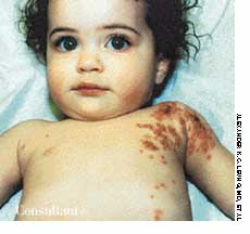
The pigmented hairy nevus that covers the entire right upper arm of this 18-month-old boy has been present since birth. More than 95% of large, congenital pigmented nevi have a hairy component.