


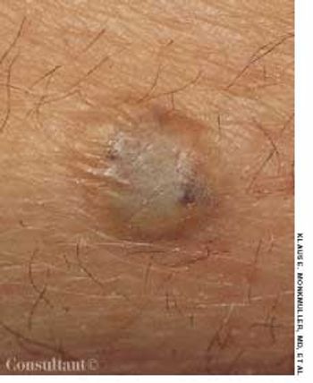
Nongenital cutaneous warts--that is, common, plantar, filiform, and flat warts--are manifestations of the human papillomavirus (HPV). These warts are among the most common dermatologic complaints seen in primary care practices and are among the most common lesions treated by dermatologists.
Dermatology
Latest News
Advertisement
Advertisement
A nonhealing ulcer recently developedin a painful facial rash that hadworsened over several months. The44-year-old patient is a heavy drinkerwith a history of elevated liver functionlevels. She has had numerousunprotected sexual contacts over theyears.
The last naturally occurringcase of smallpox was reportedin Somalia in October 1977.Despite the eradication ofsmallpox, the causative agent,variola virus, remains in existence.1,2
A pruritic facial eruption; asymptomatic, erythematous lesions;a persistent papule--can you identify the disorders pictured here?
A 5-year-old boy, who lives on a farm and routinely plays with his pet dogs, presented with these scaly, inflamed macules with a central clearingon the abdomen and forehead.
An 11-year-old boy who was receiving continuous ambulatory peritoneal dialysis because of end-stage renal disease secondary to membranoproliferative glomerulonephritis was hospitalized with hypocalcemia 2 days after subtotal parathyroidectomy. Before the surgery, multiple lesions were noted on the child's thighs (shown here), upper arms, and abdomen. The lesions, some with calcium deposits, were hard and painful. A biopsy of the lesions revealed histologic findings consistent with cutaneous calcinosis.
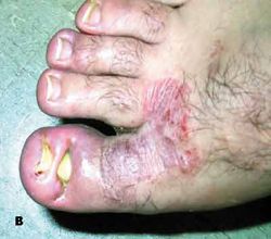
A simple trick for preventing recurrence of an ingrown toenail.
With the onset of winter, dry and scaly skin accompanied by pruritus prompted this patient to seek medical attention.
I am concerned about Dr Benjamin Barankin's recent "Photoclinic" case of an alleged brown recluse spider bite.
Erythematous patches and papules on the knuckles; a slowly enlarging papule on a finger; a painful ulcer on the thumb; rough, thickened skin around the metacarpophalangeal joints; symmetric, soft, tan papules....
Generalized weakness and malaise have bothered a 44-year-old woman for a few days. The patient has several large, flat facial lesions that have been present for years; she has never consulted a physician about them.
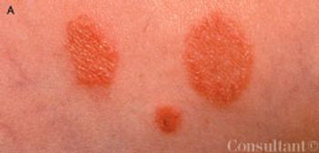
An asymptomatic acneiform eruption; persistent, mildly pruritic papules; a pustular rash that resists antibiotics--can you identify the disorders pictured here?
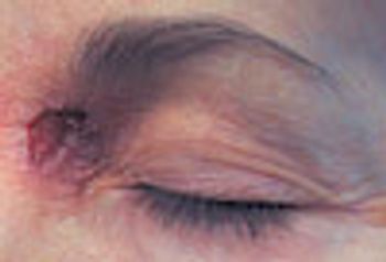
A 50-year-old woman was concerned about a nonhealing, painful lesion on the medial aspect of the left side of the nasal bridge. The lesion had been present for several weeks. The patient believed that a "cyst" had developed in the area. She had been attempting to remove it manually.
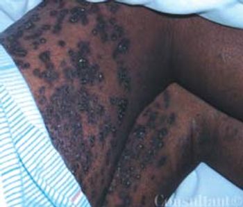
A 49-year-old man presented to the emergency department with hematemesis and 2 episodes of melena. Examination findings included resting tachycardia and melenic stool. Blood pressure was 95/50 mm Hg. Multiple raised, soft, bluish 0.3 to 1 cm lesions were noted on the trunk and extremities.
Telltale skin lesions of syphilis, gonorrhea, human papillomavirus infection, and Haemophilus ducreyi infection.
Telltale skin lesions of syphilis, gonorrhea, human papillomavirus infection, and Haemophilus ducreyi infection.
Telltale skin lesions of syphilis, gonorrhea, human papillomavirus infection, and Haemophilus ducreyi infection.
Telltale skin lesions of syphilis, gonorrhea, human papillomavirus infection, and Haemophilus ducreyi infection.
Telltale skin lesions of syphilis, gonorrhea, human papillomavirus infection, and Haemophilus ducreyi infection.
Telltale skin lesions of syphilis, gonorrhea, human papillomavirus infection, and Haemophilus ducreyi infection.
Researchers in the Netherlands investigating the relative risks of common infections in patients with type 1 or type 2 diabetes mellitus (DM1 or DM2, respectively) determined that both groups are at increased risk for lower respiratory tract infection, urinary tract infection, and skin and mucous membrane infection.
Abstract: In addition to causing pulmonary disease, infection with Mycobacterium tuberculosis can result in a wide range of extrapulmonary manifestations, including abdominal involvement. Patients with acute tuberculous peritonitis typically present with fever, weight loss, night sweats, and abdominal pain and swelling. Intestinal tuberculosis is characterized by weight loss, anorexia, and abdominal pain (usually in the right lower quadrant). A palpable abdominal mass may be present. Patients with primary hepatic tuberculosis may have a hard, nodular liver or recurrent jaundice. The workup may involve tuberculin skin testing, imaging studies, fine-needle aspiration, colonoscopy, and peritoneal biopsy. Percutaneous liver biopsy and laparoscopy are the main methods of diagnosing primary hepatic tuberculosis. Treatment includes antituberculosis drug therapy and, in some cases, surgery. (J Respir Dis. 2005;26(11):485-488)
Telltale skin lesions of syphilis, gonorrhea, human papillomavirus infection, and Haemophilus ducreyi infection.
A 35-year-old man with type 1 diabetes has had an asymptomatic rash on the lower extremities for the past several months. He denies trauma and recent illness. He has tried multiple "home remedies," but the rash has persisted. He smokes and drinks alcoholic beverages occasionally.
Progressive cough and dyspnea of 2 months' duration prompted a 23-year-old man to seek medical attention for the fourth time. On previous emergency department visits, he had received antibiotics, which failed to relieve his symptoms.
Advertisement
Advertisement
Trending on Patient Care Online
1
Cognitive Rehabilitation Linked to Functional Gains in Long COVID Trial
2
Artificial Intelligence ECG Model Identifies Patients at Higher Risk for Sudden Cardiac Death
3
FDA Authorizes Modified Risk Claim for ZYN Nicotine Pouches
4
Telehealth Mindfulness Program Linked to Sustained Low Back Pain Improvements
5