


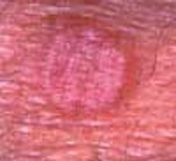
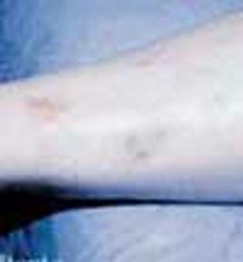
A new lesion recently arose on the right flexor forearm of a 67-year-old man. The 1-cm, pruritic, pink, circular, slightly raised lesion was perfectly homogeneous with no central clearing.
A new lesion recently arose on the right flexor forearm of a 67-year-old man. The 1-cm, pruritic, pink, circular, slightly raised lesion was perfectly homogeneous with no central clearing.
After 6 months of suffering with an infection on her finger and several failed courses of antibiotic therapy, a 53-year-old woman sought a second opinion.
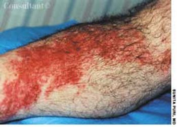
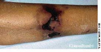
A 60-year-old man presented with redness, swelling, and pain on his right lower leg of 3 day's duration. He recalled being scratched by underbrush while hiking in the woods a few days earlier; the patient denied other recent trauma or insect bites.
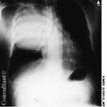
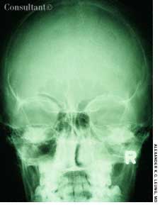
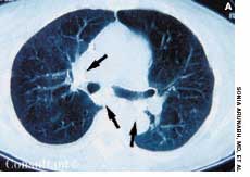
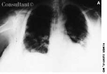
A 35-year-old man, a smoker, had right pleuritic pain, productive cough, and fever for 3 days. His pulse rate was 107 beats per minute; respiratory rate, 14 breaths per minute; blood pressure, 136/80 mm Hg; and temperature, 37.7°C (99.9°F). There were signs of right upper lobe consolidation. Laboratory studies showed hyponatremia. Chest films showed a homogeneous density in the right upper lobe.
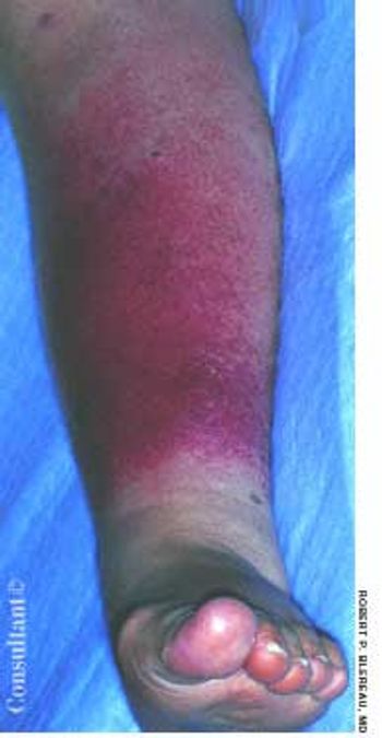
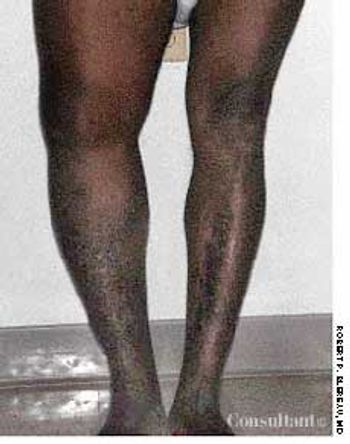
A 71-year-old man, who had recently returned from a month in Europe, complained of left lower leg swelling and pain of 1-week's duration. For many years, this obese patient had chronic venous insufficiency of both legs and chronic osteoarthritis of the knees that severely limited his ability to walk. The patient was admitted to the hospital with extensive cellulitis of the left lower leg.
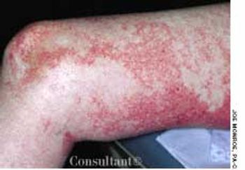
For 6 years, a 32-year-old man had a recurring rash on his back, shoulders, and chest. He stated that the rash appears in the spring, itches, and enlarges into ringlike areas. Previously, when treated with cephalexin, the rash had cleared within several weeks. Antifungal medication (econazole cream and oral terbinafine) had failed to resolve the rash.
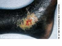
A 61-year-old woman who was receiving dialysis for diabetes-associated end-stage renal disease was hospitalized for care of an abdominal wound that had been debrided and closed. At this time, the patient had several large, indurated, red plaques with central, stellate, black eschars on her abdomen, left buttock, and legs. An early focus of ulceration was noted superior to the stapled incision.

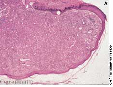
A nonpainful bump had been enlarging slowly on a 29-year-old woman's left upper eyelid. The patient wanted it removed for cosmetic reasons.
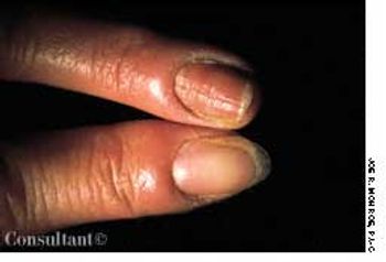
A 55-year-old-man complained of joint stiffness and red, mildly tender plaques on his fingers. He had recently sustained a trauma to the hand while at his job as a fish handler. The condition was diagnosed as erysipeloid-a skin infection caused by the gram-positive bacillus Erysipelothrix rhusiopathiae.
A 41-year-old man presented with a 3-month history of itchy, scaly feet and right hand. The left hand was unaffected.
Two days of pain in his right leg, which had been swollen for a week, brought this 69-year-old man with type II diabetes to the emergency department (ED). Three months earlier, the patient had undergone a radical retropubic prostatectomy with bilateral pelvic lymph node dissection. Examination in the ED revealed an edematous right leg indurated with a leathery-appearing thigh that was hot to the touch. His temperature was 38.7°C (101.7°F), and his white blood cell count was 21,290/µL with a shift to the left.
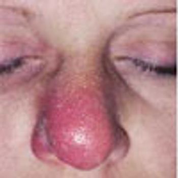
The nasal cellulitis that affects this 39-year-old woman began as right intranasal folliculitis. Because the patient was sensitive to many antibiotics, oral ciprofloxacin was prescribed.
This is a multisystem disorder characterized by oral and genital aphthae. Other symptoms include a myriad of cutaneous findings; variable systemic features include uveitis, synovitis, meningoencephalitis, and large- and smaller-vessel vascular disease.
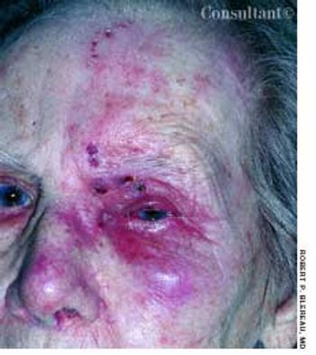
Periorbital, forehead, and nasal erythema, crusting, and pain typical of herpes zoster affected a 90-year-old woman. Reactivation of the latent varicella zoster is more common in the elderly and is attributed to impaired immunologic mechanisms.
A 49-year-old woman, severely obese but otherwise healthy, appeared for a preemployment medical examination. She neither smoked cigarettes nor drank alcohol. She had no respiratory problems and recalled no family history of such. A baseline mammogram taken 4 years earlier showed no abnormalities, and the patient was not under care for any medical condition. Results of physical examination were normal, except for the obesity-which made it difficult to determine breast masses with confidence.
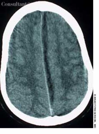
For 7 days, a 10-year-old boy had had a headache and a fever (temperature, 38.8°C [102°F]); a viral upper respiratory tract infection had been diagnosed. His parents brought him to the emergency department when weakness in his right leg developed, which impaired walking.
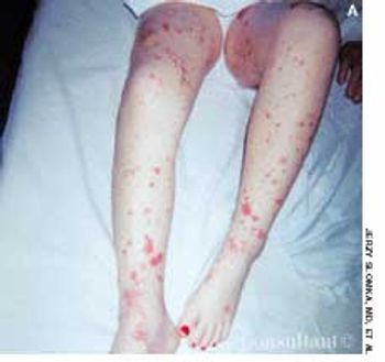
A 6-year-old girl was evaluated in the emergency department for a leg rash that had spread to the buttocks. The lesions first appeared earlier in the day and worsened hourly. The child's mother reported that her daughter was in good health until a low-grade fever, nonproductive cough, sore throat, and headache developed 5 days earlier. The youngster also complained of neck pain with movement.
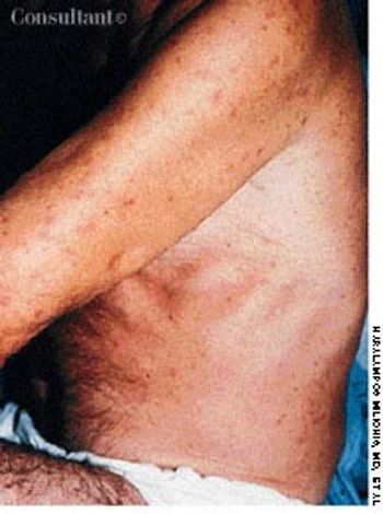
A 49-year-old farmer was hospitalized because of a 3-week history of intermittent fever, fatigue, anorexia, generalized myalgias, and malodorous sweating. A nonpruritic, nonhemorrhagic, maculopapular rash recently had developed on his arms, legs, and trunk. The reddish lesions were less than 1 cm in diameter. There was no history of antibiotic or antipyretic drug therapy, and no abnormalities were found on physical examination.

Ten days before presenting for evaluation, a 69-year-old man began to experience neuralgic pain and noticed the eruption of painful erythematous macules and papules on the right side of his chest. Within 24 to 72 hours, vesicles and pustules arose at the site. One week after onset, several of the lesions dried and crusted.
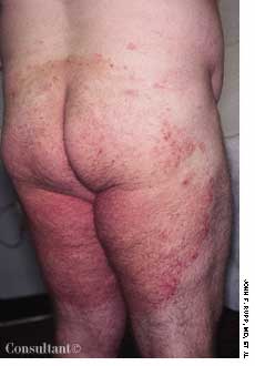
A 44-year-old woman had a painful, burning rash for 4 months. The erythematous eruption was evident on the thighs, fingers, buttocks, abdomen, and perineal and intergluteal areas. Application of triamcinolone cream and emollients offered no relief.
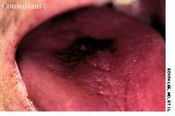
A 35-year-old HIV-positive man had a thick, black, otherwise asymptomatic patch on the top of his tongue. He did not have diabetes.
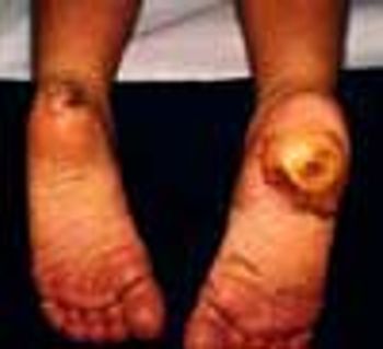
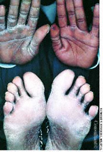
A 6-year-old girl presented with a huge ulcer on her right heel, seen here, that began as a minor laceration when she stepped on a rock several months earlier. On the left heel, there was a similar lesion in the process of healing that had also followed a minor injury. Her feet and hands were dry and hyperkeratotic.
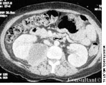
A 49-year-old woman with a history of alcoholic cirrhosis, esophageal varices, coronary artery disease, diabetes mellitus, and hypertension presented to the emergency department with a 2-day history of fever, chills, nausea, and back and abdominal pain. The pain began on the right side, progressed to the lower back, and radiated into the right anterior thigh and groin area.
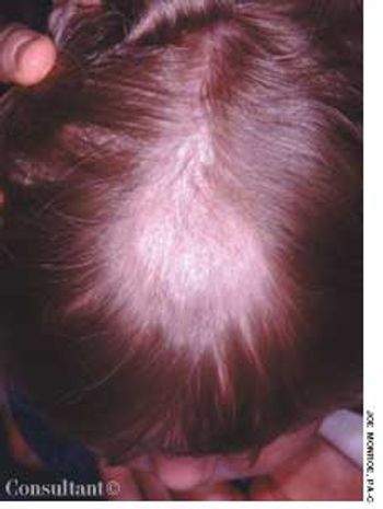
The parents of a 3-year-old girl sought evaluation of their daughter's hair loss. During the past several months, a large patch of alopecia with scaling had developed. The differential diagnosis included seborrhea, trichotillomania, and tinea capitis.

A 3-month-old female infant presented with a mass in the umbilical area. During the neonatal period, an infection of the umbilical cord had resulted in the formation of exuberant granulation tissue at the base of the umbilicus.