Articles by Victoria P. Werth, MD
This is a multisystem disorder characterized by oral and genital aphthae. Other symptoms include a myriad of cutaneous findings; variable systemic features include uveitis, synovitis, meningoencephalitis, and large- and smaller-vessel vascular disease.
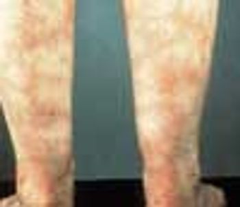
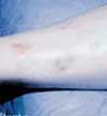
Livedo reticularis is the cutaneous manifestation most frequently associated with the antiphospholipid syndrome (APS). It manifests as a dusky, violaceous vascular discoloration with a reticulated pattern on the upper and lower extremities.
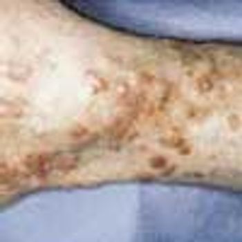
Erythema elevatum diutinum (EED) is a rare and usually chronic form of vasculitis. It has been postulated that EED is the result of immune complex formation in small vessels secondary to some antigenic stimulation.
A variety of rheumatic diseases-systemic lupus erythematosus (SLE), rheumatoid arthritis (RA), and the vasculitides among them-manifest as “lumps, bumps, and holes” involving the extremities. Each of these diseases works through specific mechanisms on different structures of the skin to produce a distinctive pathology. In doing so, each provides clues to the cause, which the history and physical examination can help confirm.
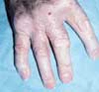
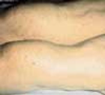
Various types of cutaneous lesions may occur in association with RA, including rheumatoid nodules, rheumatoid neutrophilic dermatitis, vasculitis, palpable purpura, and pyoderma gangrenosum. Many of these manifestations-including rheumatoid nodules-are specific for RA. The presence of these nodules is associated with seropositive disease and with a more severe, erosive clinical presentation. The nodules appear in 20% to 30% of patients with RA. Sites of predilection are those subject to shear stress, including the subcutaneous tissues over the extensor aspects of the elbow region, over the sacrum in bedridden persons, and at the pericardial and pleural surfaces.
Cutaneous and subcutaneous lesions of the extremities may be clues to the presence of rheumatic diseases such as systemic lupus erythematosus (SLE), rheumatoid arthritis (RA), and the vasculitides. The history and physical examination can generally help confirm the cause. Skin biopsy is sometimes necessary for a definitive diagnosis; useful results depend on a technique that gives the depth necessary to see the pathology and proper interpretation of biopsy specimens by an experienced dermatopathologist.

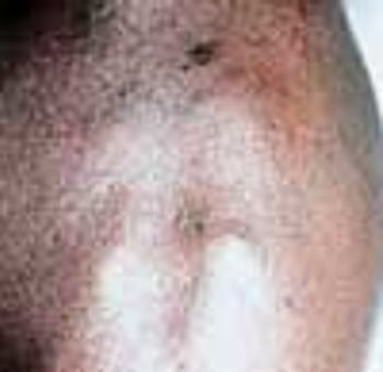
Pyoderma gangrenosum (PG) is a chronic, recurrent condition characterized by cutaneous ulceration. In half of patients, PG is associated with an underlying illness, such as inflammatory bowel disease, RA, SLE, or a lymphoproliferative disorder.


