Dermatology

Latest News


CME Content


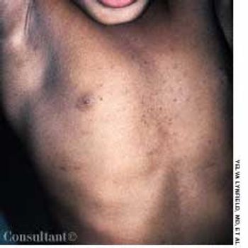
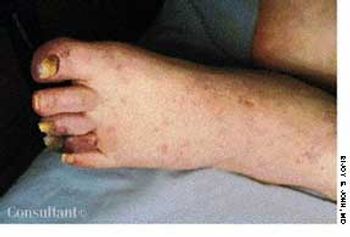
A 65-year-old man presented with bluish toes and petechiae on his toes and feet 3 days after having undergone renal angiography. Bilateral pitting pedal edema was noted, and laboratory tests revealed proteinuria, eosinophilia, and an erythrocyte sedimentation rate (ESR) of 65 mm/h.

This 27-year-old man complained that a facial rash of several years' duration had worsened during the past few months. Hypopigmented macules with scale were especially prominent on the eyebrows and in the nasolabial folds; a moderate amount of scale was noted on the scalp. The patient was seropositive for HIV.

A 56-year-old man was admitted to the hospital with right lower lobe pneumonia, which was exacerbated by smoking-induced chronic obstructive pulmonary disease (COPD).

An 80-year-old man with a history of congestive heart failure, coronary artery disease, cardiomyopathy, and thoracic and abdominal aneurysms was taken to the emergency department because of mental status changes, back pain, and ecchymotic areas over his body. The ecchymoses started on his back 5 days before admission and spread to his abdomen.

Tan-pink acneiform lesions on the face of a 15-year-old girl had not responded to topical acne therapy. A 1 × 0.5-cm, elevated subcutaneous nodule was noted on the right lateral knee. The lesions on her face and knee had been present for 11 years. The family history was noncontributory.

A 35-year-old man had a 5-year history of progressive hair loss characterized by follicular inflammation with destruction of the follicle and consequent permanent alopecia. Almost the entire scalp was involved. A few pustules were seen on examination, but the clinical picture was mostly one of scarring and irreversible hair loss.

A 79-year-old nursing home resident was hospitalized for evaluation of hyperkalemia and leukocytosis. Her medical history included hypertension, respiratory failure with subsequent tracheostomy placement and ventilator dependency, and anemia. Both of her legs had been amputated above the knee secondary to complications of type 2 diabetes mellitus.

After 3 months of seeing this painless mass at the angle of the 3-year-old's left jaw, his parents sought medical advice for their son. The youngster had no constitutional symptoms. A Mantoux test was performed, and an erythematous, indurated area measuring 15 mm in diameter was found at the test site 48 hours later.

The pigmented hairy nevus that covers the entire right upper arm of this 18-month-old boy has been present since birth. More than 95% of large, congenital pigmented nevi have a hairy component.

This erythematous, papulovesicular eruption developed in a hospitalized patient who had a febrile illness that was accompanied by drenching sweats. Miliaria rubra, or prickly heat, is caused by sweat trapped in obstructed eccrine glands of the epidermis. Inflammation, irritation, and pruritus can occur along with the rash.

An otherwise healthy 34-year-old woman was concerned because of the abrupt onset of rapid hair loss, accompanied by scaling of the underlying skin. The disorder had begun 3 months earlier, and the right parietal and temporal areas were now red and swollen and had adherent scale. An antinuclear antigen titer was negative. Biopsy revealed changes consistent with lichen planus of the scalp, also known as lichen planopilaris and lichen planus follicularis.

A mildly itchy, tender “red bump” on his eyelid concerned a 68-year-old man. Two years earlier, a basal cell carcinoma, which started as a “red bump,” had been removed from his cheek. This lesion had been present for 2 weeks; the patient noted a small amount of discharge at the site in the mornings.

A 21-year-old woman had suffered recurrent nosebleeds and pain in her nose for the previous 2 months. Physical examination revealed an extremely vascular, slowly enlarging intranasal growth on the anterior surface of the septum.

A 10-year-old boy presented with a 6-month history of a painless mass on the left side of the scrotum. The overlying skin had a bluish discoloration. The mass felt like a “bag of worms.” When the boy stood, venous varicosity could be palpated along the spermatic cord. This venous distention increased when he performed Valsalva's maneuver and decreased when he was recumbent.

A 76-year-old man reported a 3-month history of an asymptomatic, raised, reddened lesion on his penis. The patient had type 2 diabetes mellitus. In 1994, a basal cell carcinoma had been excised from his chest and, 3 years later, a squamous cell carcinoma was excised from his left temple.