


A 59-year-old woman comes to your office for evaluation of her heart murmur.During the last several months, she has tired more easily and has had less energy.Recreational activities, such as lap swimming, have become difficult becauseshe is easily winded. She denies chest pain, foot swelling, and nocturnal dyspnea.
Cardiology
Latest News
Advertisement
Advertisement
A 36-year-old man with a 15-year history of episodic migraine presents to the emergency department (ED) at 5 AM witha right-sided throbbing headache of 4 hours' duration. The headache awakened him, which is typical of his more severemigraine attacks. Unfortunately, the patient forgot to refill his prescription for pain medication and did not "catch" thisheadache in time. He took an over-the-counter combination of aspirin and caffeine, which seemed to help for about 60minutes, but the headache has returned full force. He has vomited twice-another characteristic typical of his migraineattacks
A 3-year Australian study found that when patients who underwent bypass surgerywere given coenzyme Q10 for a week or more before the operation, their heartmuscle tolerated stress better, recovered more quickly, and had better pumpingability after surgery than did the heart muscle in patients given placebo.1
Many of the approximately 12.4 million Americans whohave cardiovascular disease are not being treated with themost up-to-date risk-reduction therapies.1 Strong evidencefrom recent studies, such as the National Heart, Lung, andBlood Institute’s Adult Treatment Panel III report2 and theHeart Outcomes Prevention Evaluation (HOPE) trial,3demonstrate the need for more aggressive use of appropriatemedical and lifestyle therapies for these patients.
Q:What is the best and most efficient method ofevaluating pulmonary function in primary careoffice practice?
A 49-year-old man presents for a routine examination. He has a 15-year history of essential hypertension and a 7-year history of hypercholesterolemia and type 2 diabetes mellitus.
A 64-year-old man has experienced melena 4 or 5 times in the previous24 hours. He has had no emesis but complains of moderate epigastric pain.He has had similar-although milder-pain in recent weeks.
I wish to add ileus to the list of atypical presenting symptomsof acute myocardial infarction (MI) in an article byDrs William J. Brady, Jr, Andrew D. Perron, and Chris A.Ghaemmaghami (CONSULTANT, July 2001, page 1153).
What are the current recommendations for hormonal contraception (eg, oralcontraceptives, medroxyprogesterone injections) in patients with migraine?
My patient is an 87-year-old woman who requires transfusion every 3 weeksfor chronic GI bleeding.
A 36-year-old man who had collapsedand sustained a bruised right shoulderwas brought to the emergency departmentwith acute emesis, cephalgia,blurred vision, aphasia, and righthemiparesis. He was confused but ableto follow simple commands.
35-year-old woman presents to the emergency department with palpitations and chest pain. She is aware of her heart beating fast but not irregularly. She notes associated discomfort in the center of her chest.
22-year-old man is hospitalized because of severe hypertension. In addition, he reports exertional retrosternal pressurelike pain, with a severity of 5/10, that lasts for 2 to 3 minutes.
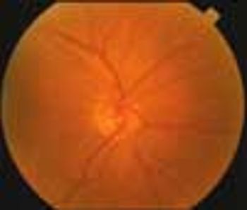
A 71-year-old man presented with a 6-week history of decreased vision in his right eye. The patient, who had hypertension and migraine headaches, had successfully recovered from a stroke that occurred 1 year earlier. His medications included aspirin, 81 mg/d, clopidogrel, atenolol, and furosemide. He also took gabapentin, 300 mg hs, for his migraine headaches. He had a remote history of cigarette smoking.
The prevalence of type 2 diabetes is expected to continue to increase rapidly, and it is not surprising that the issue of the potential effects of different classes of antihypertensive drugs on glucose metabolism and glycemic control has sparked debate.
A 57-year-old woman complains of burning and dryness in her left eye and altered sensation in her mouth when eating; these symptoms began the day before. A coworker who had noticed facial asymmetry recommended that she seek medical attention.
Up to 10% of Americans older than 20 years have type 2 diabetes, and more than 20% have the metabolic syndrome. The prevalence of both diseases has risen by 33% over the past decade as a result of an increasingly sedentary lifestyle, the obesity epidemic, the growth of ethnic groups at risk for the disease, and the aging of the population.
New guidelines recommend a different regimen for patients with native valve endocarditis caused by MRSA.
Five days ago, a 46-year-old woman experienced dull, aching retrosternal pain that radiated toward the left shoulder. The pain was accompanied by diaphoresis and did not abate; she was hospitalized.
As many as half of patients who are evaluated for abdominal pain do not receive a precise diagnosis. And for about half of those who are given a diagnosis, the diagnosis is wrong. In this article, I will use actual cases (not "textbook" examples) to illustrate an approach to abdominal pain that begins with a careful differential diagnosis. I also offer some general guidelines for evaluating patients.
An 81-year-old man is seen for follow-up of leukocytosis detected during a recent hospitalization for community-acquired pneumonia. The leukocytosis had not resolved by the time he was discharged.
A systematic approach to the patient with resistant hypertension is both cost-effective and rewarding because the evaluation will probably reveal the cause. Initial considerations include lack of adherence, inappropriate treatment, drug-drug interactions, volume overload, and white-coat hypertension.
An 83-year-old man with a history of hypertension and coronary artery disease presented with a 4-day history of mental status changes, slurred speech, and difficulty ambulating. He reported a lack of appetite and weakness of several days.
A 56-year-old woman was referred for management of severe hyperlipidemia. Her family history included hypercholesterolemia and premature coronary artery disease.
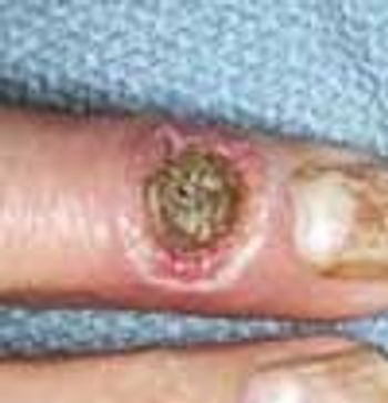
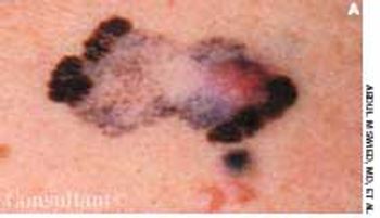
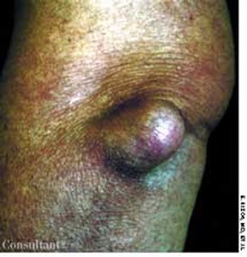
Cutaneous manifestations develop in approximately 30% of persons with diabetes. Premature atherosclerosis is a common complication of the disease that can cause peripheral infarction, ulceration, and necrosis.
Advertisement
Advertisement