Sarcoidosis in a 45-Year-Old African American Man and Rheumatoid Nodules
Skin signs of systemic disease: sarcoidosis, rheumatoid nodules, Muir-Torre syndrome, diabetic vasculopathy, hyperlipidemic nodules, zinc deficiency, and Sister Mary Joseph nodule.
Sarcoidosis

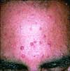
A 45-year-old African American man requested treatment of "keloids" that had developed 18 months earlier. The patient also complained of dyspnea and exertion; there was no history of trauma.
Innumerable firm, hyperpigmented papules, plaques, and nodules were noted on the patient's trunk, upper extremities, and neck. A biopsy disclosed dermal noncaseating granuloma. The angiotensin-converting enzyme level was elevated, and chest films demonstrated hilar adenopathy and parenchymal infiltrates; these findings confirmed the diagnosis of pulmonary sarcoidosis.
Sarcoidosis is a systemic granulomatous disease that involves the respiratory tract in 88% of affected patients. It is most prevalent in persons between the ages of 20 and 40 years. Northern Europeans and blacks are most commonly affected. In the United States, sarcoidosis occurs in 10.9 per 100,000 whites and 35.5 per 100,000 blacks.1
This patient takes hydroxychloroquine, 200 mg bid; systemic corticosteroids are given for infrequent exacerbations of his pulmonary symptoms. Although there is no cure for sarcoidosis, the disease can be adequately controlled in many patients with such combination therapy.
Sarcoidosis can closely mimic keloids and therefore needs to be considered in the differential diagnosis of any cutaneous eruption in African Americans.
(Case and photograph courtesy of Ted Rosen, MD.)
REFERENCE:1. Amin N. What's wrong with this picture? 26-year-old man with fever, erythematous nodules, and cough. Consultant. 2000;40:1953-1957.
Rheumatoid Nodules


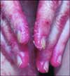
A 76-year-old woman with a 40-year history of rheumatoid arthritis (RA) had repeatedly refused treatment with disease-modifying drugs, including methotrexate. Nodules began to develop 15 years after the initial diagnosis; they recurred after surgical removal.
Rheumatoid nodules, a major diagnostic criterion for RA, are one of the most common extra-articular manifestations of the disease. The incidence of nodules among patients with RA has been reported to be 20% to 30%.1,2
Nodules can be subcutaneous or may form on any body surface, such as heart valves, lungs, and vocal cords. Subcutaneous nodules tend to develop on pressure points. The extensor surface of the elbows is the most common site. When they arise along tendon sheaths, range of motion may be limited and tendon rupture, particularly at proximal interphalangeal joints, can occur. Rheumatoid nodules can develop in bedridden patients on the occiput and ischium.
The nodules' necrotic center is encompassed by palisading fibroblasts, which are surrounded by lymphocytes that can produce IgG and IgM rheumatoid factor (RF). Most patients who have rheumatoid nodules are seropositive for RF. However, rheumatoid nodules have occurred in patients who have a negative RF assay and no evidence of arthritis.3
Rheumatoid nodules can obliterate the right ventricular cavity, which leads to right-sided heart failure.4 An association with paraneoplastic syndrome was suggested by the report of their development before the onset of B-cell lymphoma.5 Fungal colonization of pulmonary rheumatoid nodules has been reported.6 Accelerated rheumatoid nodulosis, especially that of the hands and feet, has been described in patients who are taking methotrexate for RA and in children who have juvenile RA.7,8 Rheumatoid nodules have developed in patients following leflunomide therapy.9
Surgical removal of the nodules is reserved for patients with local pain, nerve compression, limited range of motion, infection, or erosion.10 Surgically removed nodules may recur.
(Case and photographs courtesy of Ildiko Lingvay, MD, and KoKo Aung, MD.)
REFERENCES:
1. Yoshida M, Belt EA, Kaarela K, et al. Prevalence of mutilans-like hand deformities in patients with seropositive rheumatoid arthritis. A prospective 20-year study. Scand J Rheumatol. 1999;28:38-40.
2. Vlak T, Grazio S, Jajic Z. Occurrence of subcutaneous rheumatoid nodules in patients with rheumatoid arthritis in Croatia. Reumatizam. 1998;46:21-25.
3. Hewitt D, Cole J. Rheumatoid nodules without arthritis. Australas J Dermatol. 2005;46:93-96.
4. Abbas A, Byrd BF 3rd. Right-sided heart failure due to right ventricular cavity obliteration by rheumatoid nodules. Am J Cardiol. 2000;86:711-712.
5. Courtney PA, Wright GD, Finch MB. "Rheumatoid nodules" and lymphoma! Ann Rheum Dis. 1998;57:578-579.
6. Robinson C, Singh N, Addis B. Eosinophilic pneumonia and Aspergillus colonization of rheumatoid nodules. Histopathology. 2005;46:709-710.
7. Williams FM, Cohen PR, Arnett FC. Accelerated cutaneous nodulosis during methotrexate therapy in a patient with rheumatoid arthritis. J Am Acad Dermatol. 1998;39(2 pt 2):359-362.
8. Muzaffer MA, Schneider R, Cameron BJ, et al. Accelerated nodulosis during methotrexate therapy for juvenile rheumatoid arthritis. J Pediatr. 1996;128 (5 pt 1):698-700.
9. Braun MG, Van Rhee R, Becker-Capeller D. [Development and/or increase of rheumatoid nodules in RA patients following leflunomide therapy.] Z Rheumatol. 2004;63:84-87.
10. Arnold C. The management of rheumatoid nodules. Am J Orthop. 1996;25:706-708.
Muir-Torre Syndrome

Asymptomatic facial and truncal papules had begun developing several years before this 55-year-old man sought medical care. The lesions were slightly yellowish or reddish, and many had a central punctum. Biopsy revealed a microscopic picture consistent with sebaceous adenoma.
Colon cancer was a significant factor in the patient's history: he had undergone resection 5 years earlier, and the disease had been diagnosed in his brother, sister, and niece. Moreover, his brother had similar facial papules. Thus, the diagnosis of Muir-Torre syndrome was made. This rare autosomal dominant disorder is characterized by multiple sebaceous tumors (including adenomas, adenocarcinomas, and epitheliomas) in association with multiple adenocarcinomas of the colon. The patient refused the option of removal of any papules.
(Case and photograph courtesy of Reynold C. Wong, MD.)
Diabetic Vasculopathy

Cutaneous manifestations develop in approximately 30% of persons with diabetes. Premature atherosclerosis is a common complication of the disease that can cause peripheral infarction, ulceration, and necrosis.
As seen here on the finger of a 64-year-old man who has a 25-year history of diabetes mellitus, ulceration secondary to insensible trauma can arise from diabetic neuropathy. Healing of such a lesion may be further complicated by vascular insufficiency related to the disease.
The pathogenic mechanism of vascular disease in diabetes is not clearly understood. Early diagnosis and tight glucose control afford the best opportunity to delay disease progression.
This patient was referred to a vascular surgeon for evaluation and treatment. *
(Case and photograph courtesy of Charles E. Crutchfield III, MD, Eric J. Lewis, MD, and Humberto Gallego, MD.)
Hyperlipidemic Nodules


A 56-year-old man was admitted to the hospital with right lower lobe pneumonia, which was exacerbated by smoking-induced chronic obstructive pulmonary disease (COPD).
The patient's hydration status was good. The examination and chest film revealed evidence of right lower lobe consolidation. His pulse rate was 100 beats per minute; temperature, 38.2°C (100.8°F); and respiration rate, 30 breaths per minute. The laboratory workup yielded the following levels: total triglycerides, 1800 mg/dL; total cholesterol, 860 mg/dL; high-density lipoprotein, 55 mg/dL; and low-density lipoprotein, 240 mg/dL.
The patient had very firm, nontender nodules overlying the tendons of both hands and the anterior shins of both legs. He reported that many members of his family, including his father and 3 brothers, had similar lumps. His father had died at age 48 and a brother at age 44--both of acute myocardial infarction. The physical and laboratory findings, combined with the family history, led to the diagnosis of familial hyperlipidemia. The nodules on the extremities were xanthomas typical of this condition.
The patient's acute COPD exacerbation secondary to pneumonia responded promptly to intravenous antibiotics, intravenous corticosteroids, oxygen, and breathing treatments. After further evaluation, an ECG showed nonspecific T-wave changes, a stress test was positive, and an angiogram revealed 3 diseased vessels. The patient underwent triple bypass surgery. A statin was prescribed for the hyperlipidemia.
(Case and photographs courtesy of Navin M. Amin, MD.)
Zinc Deficiency


A 44-year-old woman had a painful, burning rash for 4 months. The erythematous eruption was evident on the thighs, fingers, buttocks, abdomen, and perineal and intergluteal areas. Application of triamcinolone cream and emollients offered no relief.
The patient was living in a nursing home. She had a history of chronic alcoholism with subsequent liver failure but denied using alcohol for the past 3 months. She complained of poor appetite and progressive weight loss. Her medications included vitamin supplements.
The patient was cachectic and extremely weak; eye contact was poor. She showed no signs of jaundice. The extensive eruption exhibited focal annularity and no vesiculation. Fine scale was most dense along the leading edges of the lesions; results of a potassium hydroxide examination ruled out a dermatophyte infection.
Laboratory studies revealed no evidence of anemia; however, the serum zinc level was 28 µg/dL (normal, 80 to 120 µg/dL). Subsequently, it was learned that the patient's vitamin supplements did not contain zinc.
A punch biopsy specimen showed psoriasiform changes, parakeratosis, and a sparse superficial perivascular lymphohistiocytic infiltrate--skin changes that are secondary to zinc deficiency. The lack of eosinophils in the infiltrate ruled out contact dermatitis; the absence of stigmata and a family history of psoriasis excluded that disease. Zinc deficiency, or acrodermatitis enteropathica, was diagnosed.
Acrodermatitis enteropathica formerly referred to an inherited disorder that predominantly affected infants. The definition now includes any dermal syndrome that involves zinc deficiency with acral eruption.1
Alcoholism and cirrhosis cause hypozincemia by increasing urinary loss of the metal, which may already be at low levels because of insufficient dietary intake. Other causes of nonhereditary acrodermatitis enteropathica include GI diseases and catabolic processes. A daily oral dose of zinc sulfate, 200 mg, was prescribed for this patient.
One week after these pictures were taken, the patient died of cardiac arrest. The association of zinc deficiency with her death is unclear.
(Case and photographs courtesy of Joe Monroe, PA-C.)
REFERENCE:
1. Freedberg IM, Eisen AZ, Wolff K, et al, eds. Fitzpatrick's Dermatology in General Medicine. 5th ed. New York: McGraw-Hill; 1999:1738-1744.
Sister Mary Joseph Nodule

An 85-year-old man presented with intermittent abdominal pain of several years' duration. Two years earlier, he had undergone a laparoscopic cholecystectomy that was converted to an open procedure. A CT scan performed a year after surgery yielded normal findings.
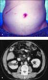
The physical examination revealed an oozing mass that had been present for 3 weeks along the periumbilical area (A). Barium swallow and endoscopic examination results were unremarkable. A CT scan demonstrated multiple abdominal masses with invasion into the pancreas (B).
A biopsy of the node disclosed areas of squamous cell carcinoma and adenocarcinoma; the periumbilical mass was a Sister Mary Joseph nodule. Immunohistochemistry stains demonstrated cytokeratin 7 and carcinoembryonic antigen. No clear primary source of the malignancy was discovered; the differential diagnosis included malignancy from the pancreas, colon, and bile duct.
Sister Mary Joseph nodule is named after Dr William Mayo's assistant who became the superintendent of Saint Mary's Hospital in Rochester, Minn, in 1892.1 During the preoperative preparation of patients, she noted that indurated nodules along the periumbilical area were associated with advanced intra-abdominal malignancy.
The nodule generally portends a poor prognosis. In one study, patients survived an average of 11 months after umbilical metastasis appeared; the nodule was the presenting symptom of malignancy in nearly 50% of patients.2 The primary tumor is identified in about 9% to 17% of nodules that metastasize to the umbilicus.3,4
Because of the extent of the disease and the poor prognosis, this patient was given palliative chemotherapy. He died 3 months later.
(Case and photographs courtesy of Manny C. Katsetos, MD, and Steven V. Angus, MD.)
REFERENCES:
1. Hill M, O'Leary JP. Vignettes in medical history. Sister Mary Joseph and her node. Am Surg. 1996;62:328-329.
2. Pieslor PC, Hefter LG. Umbilical metastasis from prostatic carcinoma--Sister Mary Joseph's nodule. Urology. 1986;27:558-559.
3. Dubreuil A, Dompmartin A, Barjot P, et al. Umbilical metastasis for Sister Mary Joseph's nodule. Int J Dermatol. 1998;37:7-13.
4. Powell FC, Cooper AJ, Massa MC, et al. Sister Mary Joseph's nodule: a clinical and histologic study. J Am Acad Dermatol. 1984;10:610-615.