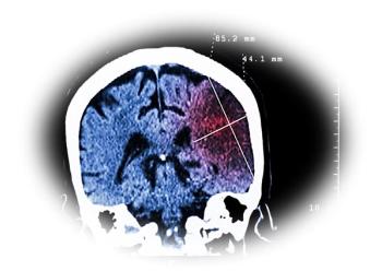
Diaphragmatic Hernia: Delayed Presentation is Common
A 48-year old man presents to the emergency departmentwith constant, dull epigastric pain and right upperquadrant pain. The pain has been present for 2 to 3months; does not radiate; has not changed its pattern; andis not associated with fever, nausea, vomiting, diarrhea, orchanges in urine or stool color. There are no alleviating orprecipitating factors.
Figure 1
Figure 2
1. Constant epigastric and right upper quadrant pain
A 48-year old man presents to the emergency departmentwith constant, dull epigastric pain and right upperquadrant pain. The pain has been present for 2 to 3months; does not radiate; has not changed its pattern; andis not associated with fever, nausea, vomiting, diarrhea, orchanges in urine or stool color. There are no alleviating orprecipitating factors.
The patient has no history of hypertension, diabetes,peptic ulcer disease, gastroesophageal reflux disease, hepatitis,or pancreatitis. He denies recent trauma but was involvedin an accident 7 months ago in which he was hit bya van while walking. He was hospitalized for a "head injury"at the time and is currently disabled as a result, buthe denies any orthopedic or abdominal injuries. He doesnot use alcohol or tobacco.
The patient has not lost weight, has no urinarysymptoms, and is currently in no acute distress. Temperatureis 36.3oC (97.4oF); heart rate, 76 beats per minute;respiration rate, 18 breaths per minute; and blood pressure,130/70 mm Hg. Results of neurologic, head, eye,ear, nose, throat, heart, and neck examinations are normal.Breath sounds are decreased at the right lung base,but lungs are otherwise clear. Bowel sounds are normal;no fecal occult blood. Deep palpation of the abdomen revealstenderness in the right upperquadrant, but there is no rebound orguarding. Murphy sign is absent.Back examination shows no costovertebralangle tenderness.
Results of a complete blood cellcount, basic metabolic panel, liverfunction tests, amylase and lipasemeasurements, and urinalysis arenormal. ECG shows normal sinusrhythm.
You order radiographs of thechest and abdomen. What findingson these films point to the cause ofthe patient's pain, and what furtherinvestigation is warranted?
Figure A
Figure B
Figure C
Figure D
Figure E
1. Constant epigastric and right upper quadrant pain:The frontal upright chest film demonstrates elevation ofthe right hemidiaphragm (A, arrow) but is otherwise unremarkable.The radiograph of the pelvis reveals fracturesof the superior left pubic bone (B, arrows).
You order a CT scan to investigate these findings further.An axial image (C) demonstrates the dependent viscerasign, in which the liver makes contact with the posteriorribs (arrow); this strongly suggests a right-sided diaphragmatichernia.
You order an MRI study to confirm your suspicions.Coronal (D) and sagittal (E) T1-weighted images show arent in the center of the diaphragm (arrows) throughwhich the right lobe of the liver herniates. The CT andMRI images clearly point to increased intra-abdominal/intrapelvic pressure as the cause of both the pelvicfractures and the rupture of the diaphragm.
Diaphragmatic injury usually results from a suddenand substantial increase in intra-abdominal pressure. Duringforced breathing, for example, the muscles of the abdominalwall contract, pushing the diaphragm upward andthe ribs inward and downward. The opposed motions cancontribute to injury, especially if a rib is fractured becauseit can tear the diaphragm. Violent, paroxysmal coughingcan also cause diaphragmatic injury. In this patient, thecause of the increased pressure was the motor vehicle accidenthe was involved in 7 months earlier. Diaphragmatichernias often present in a delayed fashion, as was the casehere; symptoms sometimes do not develop until yearsafter the inciting traumatic event.
Diaphragm injury is 3 to 5 times more likely to occuron the left side than on the right. One explanation for thisdifference is that the liver provides an extensive protectivebarrier against injury on the right side. In addition, thethickness of the diaphragm is typically greater on theright side than on the left. However, the forces involved inthis patient's accident were directed in such a way andwith such impact that a right-sided rupture resulted.
The signs and symptoms of diaphragmatic rupturecan include:
- Dyspnea.
- Dysphagia.
- Sharp epigastric pain.
- Chest pain that radiates to the ipsilateral shoulder(phrenic nerve injury).
- Bowel sounds heard in the lower and middle chest.
- Decreased breath sounds on the injured side.
Characteristic abnormalities on a chest radiographinclude:
- A mushroom-like shadow that suggests elevation of theaffected hemidiaphragm.
- A shift of the heart and mediastinal structures to the unaffectedside (present in this patient's radiographs, but toa very small degree).
Diaphragmatic rupture is rarely diagnosed, however,on the basis of radiographic findings. On a chest film, the inferiorsurface of the diaphragm is camouflaged by the organsand soft tissues of the abdomen. The characteristic abnormalitiesare often attributed to a small pleural effusionwith atelectasis or elevation of the hemidiaphragm.
CT or MRI is needed to confirm the diagnosis. An MRIscan performed in 3 planes or a helical CT scan with coronaland sagittal reformations can be used. Discontinuity of thediaphragm-with or without herniation-is apparent onsuch scans. Evidence of bowel, the stomach (typically), orsolid organs herniating through the diaphragm also indicatesa rupture.
This patient's diaphragm was surgically repaired, andhis pain resolved.
2. Severe lower abdominal pain in a young woman
A 19-year-old woman complainsof severe lower abdominal pain thatbegan 24 hours earlier. The pain,which is located mainly on both sidesof the pelvic region, occurred suddenlyand was initially sharp; it then decreasedsomewhat but has progressivelyworsened in the last few hours.The patient notes that it now hurts tomove and that the pain lessens whenshe lies still. There is no associatedfever, nausea, vomiting, diarrhea, dysuria,hematuria, or vaginal bleeding ordischarge. Her last menstrual periodended 1 week ago; she is sexuallyactive.
The patient is nulliparous; shehas no history of gynecologic, sexuallytransmitted, or other diseases, noris there any family history of gynecologicdiseases. She is currently in amonogamous relationship, and herpartner does not have any genitourinarysymptoms. She denies shortnessof breath, intrauterine foreign bodies,and changes in bowel patterns. Shedoes not use alcohol or tobacco.
Temperature is 36.8oC (98.3oF);heart rate, 80 beats per minute; respirationrate, 18 breaths per minute;and blood pressure, 100/70 mm Hg.No neck adenopathy, oral or pharyngeallesions, or petechiae are present.Heart and lungs are normal. Bowelsounds are hypoactive. The abdomenis mildly distended and tender, with guarding that isworse in both lower quadrants; no rebound. Genitalia arenormal, without discharge. Severe cervical and bilateraladnexal tenderness prevents examination for masses. Theanterior rectal wall is also tender; stool is heme-negative.
Results of a complete blood cell count, basic metabolicpanel, urinalysis, liver function tests, and amylase and lipasemeasurements are normal. A urine test for β-humanchorionic gonadotropin (β-hCG) is negative.
You order a radiograph of the abdomen. What abnormalityis evident, and what steps will you take to narrowthe differential diagnosis?
Figure A
Figure B
2. Severe lower abdominal pain in a young woman:The abdominal radiograph (A) reveals a nonspecific bowelgas pattern: prominent loops of small bowel are visibleand gas is present in the colon (arrow). Soft tissue densitywithin the pelvis extends up the pelvic side walls into thelower abdomen. This latter finding, along with the bowelgas pattern, strongly suggests free intraperitoneal fluid.
You order a CT scan of the abdomen. These imagesreveal a large area of hypodensity within the pelvis on theright side of the adnexal region (B, arrow). They alsoshow fluid within the upper abdomen (C, arrow) and alarge amount of soft tissue density surrounding the areaof hypodensity (D, arrow) that demonstrates greater attenuationthan free intraperitoneal fluid.
This constellation of findings suggests an ovarian pathology.The patient's negative urine test for β-hCG excludesectopic pregnancy. Other conditions in the differential diagnosisof ovarian pathology in a young woman include:
- Tubo-ovarian abscess. This typically has a more loculatedappearance on radiograph than is seen here. Also,a patient with a tubo-ovarian abscess usually has a feverand an elevated white blood cell count, which this womandoes not have. The patient's history of a monogamousrelationship reduces the likelihood of this diagnosis.
- Endometrioma. Patients with endometrioma often complainof pain at the time of menses that worsens over aperiod of months. However, it is relatively uncommon foran endometrioma to be accompanied by large amounts ofhemorrhage within the peritoneal cavity. The typical appearanceon CT is that of a complex cystic fluid collectionthat represents hemorrhage at different stages of evolutionas well as the ectopic endometrial tissue.
- Tumor. The presence of fat (not seen here) is diagnosticfor a teratoma (a benign lesion). However, teratomas arenot typically accompanied by acute hemorrhage. Cystadenomaand cystadenocarcinoma are both possible, althoughadenomas are far more common in this age group. Thepresence of any ovarian lesion can predispose a patient toovarian torsion.
- Functional follicular cyst. This is the most common entitythat causes hemorrhage in the pelvis of young women.These cysts are often accompanied by small amounts ofhemorrhage within the cyst or in the cul-de-sac, but occasionallythey are accompanied by a large amount of hemorrhage.These lesions also predispose patients to ovariantorsion.
The patient underwent exploratory laparotomy withevacuation of hemoperitoneum, and a right ovarian cystwas identified. The ruptured cyst was oversewn. Althoughshe required a transfusion, the patient's postoperativecourse was uncomplicated, and she was discharged after2 days.
Newsletter
Enhance your clinical practice with the Patient Care newsletter, offering the latest evidence-based guidelines, diagnostic insights, and treatment strategies for primary care physicians.


