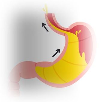
Bezoar Secondary to Diabetic Gastroparesis
A 26-year-old woman with a long history of type I diabetes mellitus (DM) presents to the emergency department (ED) with 6 days of severe nausea and anorexia. There has been no vomiting, abdominal pain, diarrhea, dysuria, fever, or other symptoms. She has no other prior medical or surgical history and was taking only insulin until her doctor recently started her on compazine and then phenergan, neither of which has helped relieve her nausea.
On physical exam she is in moderate acute distress with normal vital signs except for mild tachycardia, holding an empty emesis bag and occasionally having dry heaves. Her head and neck exam are normal except for a slightly dry appearing tongue. Her abdominal exam is benign with no palpable mass, tenderness, rebound, or guarding.
Blood is sent to the lab to ascertain a possible cause for the nausea such as hyponatremia, renal failure, ketoacidosis, infection, or pregnancy. A CBC, Chem-12, UAma and pregnancy test all are normal. Although it is typically low yield in a case like this, a KUB (kidneys, ureter, bladder) x-ray is ordered since no other cause has been found and the patient’s symptoms have shown no improvement after 6 days of conservative management, including multiple antiemetics.
The plain film of the abdomen is shown in the Figure, above right; click image to enlarge.
What pathologic finding is noted is noted on the KUB?
(Hint: there are 12 foreign bodies noted on this film, two are inside the patient.)
Answer: Bezoar secondary to diabetic gastroparesis.
Note the slightly hyperdense, somehwhat circular mass in the left upper quadrant just below the bra wire. This is a vegetable matter bezoar. An IUD is incidentally seen in the pelvis but is not related to her symptoms (nor are her 2 bra wires and 8 bra clasps). The bowel gas pattern is nonobstructive.
Discussion
Gastroparesis is most commonly caused by diabetes but can also be caused by electrolyte and endocrine abnormalities as well as medications (see Table below). Symptoms may initially include bloating, early satiety, and nausea. More severe symptoms include abdominal pain and vomiting, often 1to 2 hours after a meal. Physical exam may or may not include upper abdominal tenderness and/or signs of dehydration.
The diagnosis of gastroparesis is often initially one of exclusion with other, more serious causes of pain sometimes requiring imaging and/or endoscopy to rule them out. A gastric emptying study, however, is the diagnostic test of choice. Complications of gastroparesis may include malnutrition, dehydration, and bezoar formation, the latter two of which were seen in this case.
Gastroparesis is often a chronic recurrent condition. Treatment of gastroparesis should be aimed at correcting or controlling any complications or identifiable underlying cause, but may also include certain dietary restrictions as well as promotility agents and medications to treat nausea (see Table). Surgical treatment is a last resort when other modalities fail.
(Click on image for details on "Quick Essentials")
Newsletter
Enhance your clinical practice with the Patient Care newsletter, offering the latest evidence-based guidelines, diagnostic insights, and treatment strategies for primary care physicians.


