


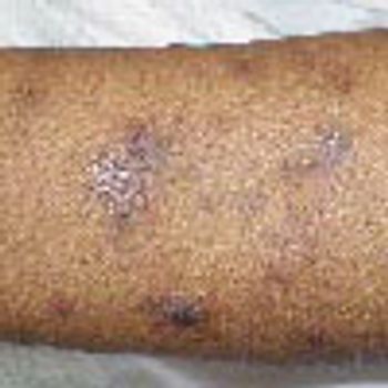
A 38-year-old African American man with HIV infection presents with numerous dyspigmented macules and patches on the extremities, abdomen, and chest; the lesions are smooth, nontender, and minimally pruritic.
Dermatology
Latest News
Advertisement
Advertisement
ABSTRACT: Risk factors for oral cancer include tobacco use and alcohol intake (especially in conjunction with tobacco use). Many benign conditions may be confused with squamous cell carcinoma, the most common type of intraoral neoplasm. Any red and/or white lesion that has surface corrugation, stippling, or induration is considered dysplastic or neoplastic until proved otherwise. Even without these clinical signs, white plaques of any size that persist for several months may represent dysplasia. These lesions should be assessed by biopsy. Risk factors for lichen planus include stress, exposure to certain foods and medications, and systemic illness. Erosive lichen planus may cause significant pain and oral dysfunction.
ABSTRACT: Only a small number of patients with celiac disease exhibit the textbook symptoms of malabsorptive diarrhea with steatorrhea, weight loss, and nutritional deficiencies. Others may present with a subclinical enteropathy, GI complaints without constitutional symptoms, persistent travelers' diarrhea, or extraintestinal manifestations alone. Be alert for suggestive signs, such as weight loss, skin lesions, oral aphthae, muscle atrophy, de-enamelization of the teeth, and vague GI symptoms, such as bloating. Helpful serologic tests include IgG and IgA antigliadin antibodies, enzyme tissue transglutaminase antibodies, antiendomysial antibodies, and total IgA. Typical endoscopic findings are mucosal atrophy, fissuring, and scalloping. In addition to a gluten-free diet, management encompasses repletion of vitamins and minerals, including iron, folate, calcium, and vitamin D; screening for thyroid disease and diabetes mellitus; bone densitometry and age-appropriate cancer screening; and pneumococcal vaccination.
Generalized papular, erythematous, nonpruritic, hyperpigmented lesions had appeared on the face, arms, chest, and abdomen of a 25-year-old homosexual man with AIDS during the previous month. Anupama Ravi, MD, of Atlanta also noted purple-red, nodular lesions in the right conjunctiva and oral cavity, especially the lower gingiva. Other pertinent physical findings included facial edema and hepatosplenomegaly.
An 89-year-old woman is seen for a rash on her back of 2 days' duration. Has Broca aphasia and dense right hemiparesis from an old stroke; remains highly communicative without words, and interactive, in the nursing home where she lives. Long-standing diabetes mellitus and hypertension contributed to the stroke and to marked peripheral arterial disease.
A 65-year-old man consults his primary care physician because of concern about nonpruritic yellowish lesions on his eyelids. He says they have been present for the past few years but have recently become more numerous.
The bites of only a few spider species produce medically significant effects in humans.
An 80-year-old man has had an asymptomatic, flesh-colored swelling on his right ear for 4 to 5 months. In the center is a 1-mm white scab pointing downward from the helix. At times, the patient shaves a white spicule that grows in this crusted area. He sleeps on his right side and does not use a cell phone.
A 38-year-old man sought treatment for the intensely pruritic swellings that had arisen on his upper lip 2 weeks earlier (A). These sharply demarcated, tender, boggy, granulomatous, pustular tumefactions are kerions, write Florence Isaac, MD, of Mohammad Dossary Hospital in Saudi Arabia and Shaun Isaac, MD, of St Petersburg, Fla. The diagnosis is based on the history of acute onset, the clinical appearance of the lesions, and the demonstration of a fungus by a potassium hydroxide (KOH) preparation of loose hairs removed from the affected area and by fungal culture. In this case, the KOH preparation revealed fungal filaments, which on culture grew Microsporum canis. A pus swab test should be performed to detect any bacterial copathogen. The differential diagnosis of kerion includes impetigo and carbuncle.
A 4-year-old girl presents with a highly pruritic rash. The day before, she had been playing outdoors at her grandmother's house. No pets were present, and the patient does not recall being stung or bitten by insects. There are bushes on the grandmother's property.

Two children, one with a history of infection, the other with a history of an allergic reaction were noted to have postinflammatory hyperpigmentation.
Application of liquid nitrogen often must be repeated several times when used to treat thick seborrheic keratoses--and still may be ineffective. For an immediate and excellent cosmetic result, try liquid nitrogen for 5 seconds, followed by gentle curettage. Any pinpoint bleeding can be stopped with aluminum chloride.
For as long as he could remember, a 27-year-old man had had a recurrent eruption on the palms and sides of the fingers. The rash was characterized by intense pruritus followed by the formation of small water blisters and increased perspiration that resolved with peeling of the skin. The dorsa of the hands were unaffected. Results of a potassium-hydroxide preparation and fungal culture of skin scrapings were negative for hyphae. The thyrotropin level was normal.
Pruritic eruptions on both arms of a 12-year-old who has played outdoors all summer; a rash on the hand of a teenage baseball player . . . might sports be responsible for these lesions?

A 38-year-old man sought treatment for the intensely pruritic swellings that had arisen on his upper lip 2 weeks earlier. These sharply demarcated, tender, boggy, granulomatous pustular tumefactions are kerions.

For as long as he could remember, a 27-year-old man had had a recurrent eruption on the palms and sides of the fingers. The rash was characterized by intense pruritus followed by the formation of small water blisters and increased perspiration that resolved with peeling of the skin. The dorsa of the hands were unaffected. Results of a potassium-hydroxide preparation and fungal culture of skin scrapings were negative for hyphae. The thyrotropin level was normal.
Squamous cell carcinoma (SCC), the second most common type of skin cancer, most often occurs on the sun-exposed skin of elderly men and women. Marjolin ulcers are SCCs that result from exposure to radiation and can arise in areas of chronic injury, typically on the extremities.
Abstract: A number of factors can contribute to a delay in the diagnosis of tuberculosis in pregnant women, including the presence of nonspecific symptoms, such as fatigue and cough; extrapulmonary manifestations; and asymptomatic disease. The diagnostic evaluation is the same as for nonpregnant patients and includes tuberculin skin testing and, when indicated, chest radiography (with appropriate shielding) and acid-fast bacillus stain and culture. Antituberculous therapy during pregnancy is generally safe and effective, although streptomycin should not be used because of the risk of vestibular or auditory damage to the fetus. For patients with active tuberculosis, treatment should be initiated as soon as the diagnosis is established. The treatment of latent infection is somewhat more controversial. The timing of the initiation of therapy is based on the risk of progression to active disease. (J Respir Dis. 2006;27(8):338-347)
Breast cancer, the most common cancer in women, is the cancer that most frequently spreads to the skin. A typical presentation is carcinoma en cuirasse, recurrent metastatic breast cancer that appears as nodules on the chest.
A 56-year-old man presented tothe emergency department with ahistory of constipation, abdominalpain, and blood-stained mucusdrainage from the rectum. The patienthad no fever; vital signs werestable. A vague fullness was felt in thelower abdomen. Rectal examinationrevealed anal warts; a patulous analsphincter; and a smooth, mobile, firmmass above the dentate line.
What are the consequences-if any-of not treating tineaversicolor?
Patients with allergic rhinitis are genetically predisposed to producing specific IgE antibodies in response to environmental allergens, such as tree, grass, or ragweed pollen or cat, dog, or dust mite allergens. Patients must have symptoms suggestive of allergies and positive skin or serologic test results that correlate with their symptoms.
A 31-year-old white woman presents with an erythematous skin eruption of 2 weeks' duration that consists of papules on the cheeks and several disk-shaped papules and plaques on the back, posterior neck, and right upper anterior chest.
A painful, vesicular eruption; an asymptomatic red macule; a rapidlyspreading erythematous plaque--can you identify the disorderspictured here?

Painful, 1- to 1.5-cm macules and papules had developed on the palms and dorsal hands and wrists of a 60-year-old man 2 weeks earlier, after a deer-hunting trip. He had not seen any ticks on his skin or clothing. The lesions persisted despite self-treatment with over-the-counter topical corticosteroids. The patient had general malaise but denied fever, chills, and arthralgia. He was not taking any medications.
Advertisement
Advertisement
Trending on Patient Care Online
1
Cognitive Rehabilitation Linked to Functional Gains in Long COVID Trial
2
FDA Authorizes Modified Risk Claim for ZYN Nicotine Pouches
3
5 FDA Decisions for Primary Care to Know from June 2026
4
FDA Expands Roflumilast Cream 0.3% Approval for Plaque Psoriasis in Children Aged 2 Years and Older
5