


Which of these GI and skin lesions should you worry about most? Is rhabdomyolysis usually the result of infection? See how well you do with this week’s questions. . .
Which of these GI and skin lesions should you worry about most? Is rhabdomyolysis usually the result of infection? See how well you do with this week’s questions. . .

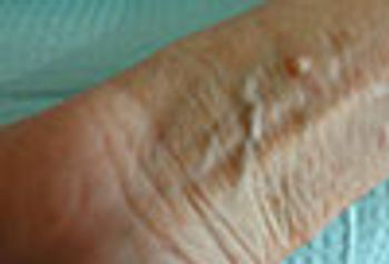
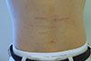
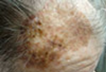
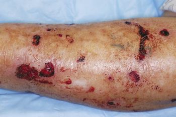
These wounds occurred when this 77-year-old woman fell when chasing her dog. Such wounds-a combination of ecchymoses and erosion caused by blunt and sheering forces on fragile skin-are common in the elderly, even after relatively mild trauma. Simple interventions will promote healing.

These classic linear, wide-mouthed, red to purple, atrophic patches are classic for stretch marks (striae distensae). The patient was in the middle of his growth spurt.
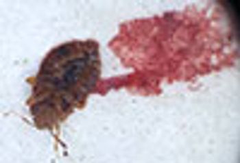
The patient assumed that the itchy rash was due to insect bites. The shape, size, color, and presence of 6 legs of the insect he captured were consistent with bedbugs and bedbug bites.
Angiofibromas and neoplasms? CRP as a marker of depression? Colorectal metastases? Can you answer all 5 of this week's questions?
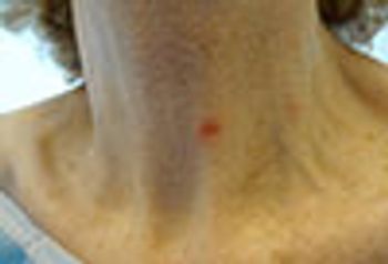
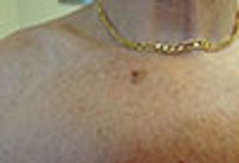
The red patch on this woman's neck is lichenoid keratosis, a variant of seborrheic keratosis. Diagnostic contenders included Bowen disease and superficial basal cell carcinoma.
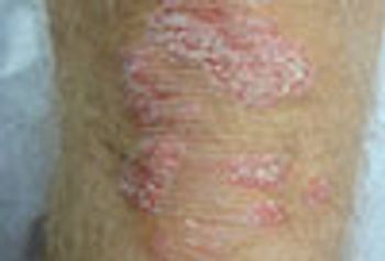
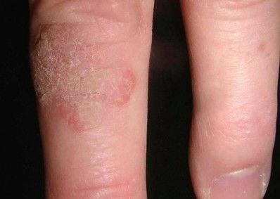
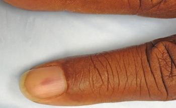
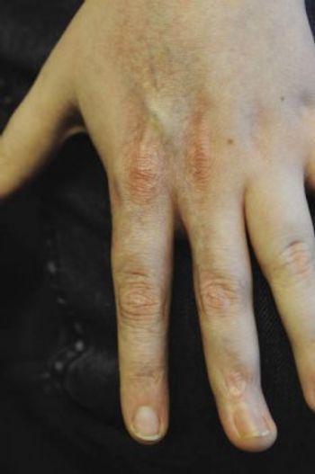
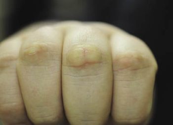
Both morphology and location favor the diagnosis of psoriasis. The presence of nail pitting, and a strongly positive family history for psoriasis, confirmed the diagnosis.

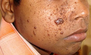
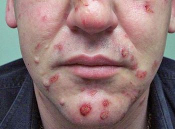
These widely scattered, dome-shaped pruritic inflammatory papules suggest insect bites. A biopsy showed mixed dermal infiltrate consisting of lymphocytes and eosinophils, consistent with an arthropod assault.
Patients often present with skin lesions that are suggestive of skin cancer. Is it or isn’t it? This week’s photo quiz tests your knowledge of several possible cancerous presentations.
Skin eruptions, MS risk, and a pain in the back: 5 new questions-your Dxs?

Spider bite? Virus? Inflammatory disease? Or, something else?
The differential diagnostic possibilities for this tender nodule included hypertrophic actinic keratosis, keratoacanthoma, squamous cell carcinoma, and verruca. The possibility of squamous cell carcinoma dictated removal with histologic examination.
A multiplicity of colors in a pigmented lesion usually dictates the need for a biopsy. In this woman with many nevi and a history of malignant melanoma, biopsy revealed an inflamed blue nevus and was also curative.
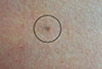
The development of eccentric pigmentation within a previously stable pigmented lesion may indicate malignant degeneration, as was the case here. Of all melanomas, lentigo maligna melanoma has the best overall prognosis.
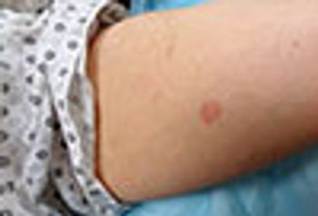
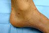
Round plaques like this one, each composed of many small erythematous papules, are classic for granuloma annulare. Large numbers of these lesions and widespread distribution is often associated with diabetes mellitus.
Finger disorders may be caused by an injury or may result from another disorder, and it’s often hard to tell what happened. This week’s photo quiz tests your ability to recognize a variety of common problems.
Chicken pox? Lupus? Impetigo? Your dx?
Rash, lumps, and bumps; extra GI IBD; and smoking: can you answer these 5 questions?

Rashes, like children, come in all shapes and sizes, and they have a variety of causes, ranging from infection to allergic reaction. This week’s photo quiz tests your knowledge of skin conditions in younger patients.
Is the nail dystrophy secondary to trauma, or something else?
This week's quiz questions challenge you to dive below the skin’s surface to come up with the correct answers.
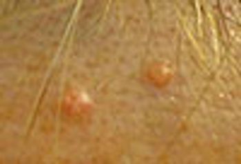
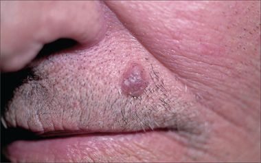
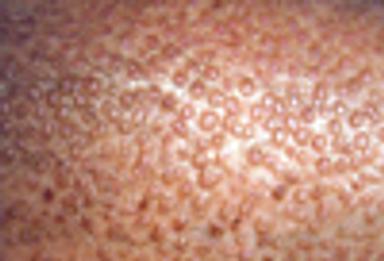
Sebaceous hyperplasia are commonly mistaken for basal cell carcinoma. These small papules with a yellowish hue and central dell is typical of the former, for which no treatment is required.
Chalk these lesions up to trauma?

This week’s questions cover a range of disorders-from infectious disease to GI problems, to HIV/AIDS. See how well you do . . .
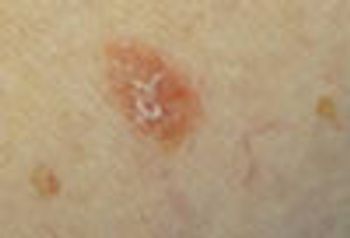
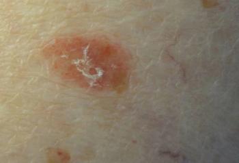
Biopsy of this red, scaly patch showed in situ squamous cell carcinoma. Treatment consisted of once-daily topical application of 5% imiquimod until erosion occurred. This off-label treatment affords an excellent cosmetic result.